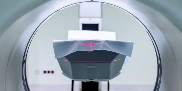
The Centre Léon Bérard, a cancer centre in Lyon and the Rhône Alpes region, has announced that it is working on three new programmes in radiotherapy. These new programs will use AI to better control the delivery of dose during therapy while reducing the duration of treatment. These projects are in the continuity of the first six already launched, also using AI in order to improve the diagnosis and monitoring of cancer treatments.
Three complementary projects
These three programs will be conducted in collaboration with a start-up TheraPanacea, an industrial company and the Creatis laboratory (CNRS UMR 5220 – Inserm U1294 – University Lyon 1 – INSA Lyon – Jean Monnet University Saint-Etienne). The Léon Bérard Center wishes to continue to use AI and new technologies to accelerate progress on the accuracy of treatments. In collaboration with the start-up Owkin, the hospital published an article showing how artificial intelligence allows doctors to improve current knowledge of mesothelioma, a rare and virulent form of cancer.
Each of the three programs will be based on three distinct but complementary technical aspects, all with the same objective: to improve patient care using tools that integrate AI. Two of these programs will intervene upstream of the treatment and the last one will be useful during the execution of the therapy.
Pre-treatment: organ delineation and MRI
The first program concerns the delineation of organs at risk, a practice called contouring. This time-consuming practice is performed by the dosimetrist and the radiation therapist. The arrival of AI could make it possible to automate this practice and thus improve its accuracy. This will be possible thanks to the automatic contouring tool Annotate, developed by TheraPanacea, whose tests carried out on dosimetry scans of more than 25,000 patients allow the contouring of more than 80 organs at risk. The tool’s accuracy is achieved through deep learning.
Alexandre Munoz, dosimetrist in the Department of Radiation Oncology at the CLB, explains that “today, when used in clinical routine, this tool is 80% faster than previous tools, and in fine, we save between 1 and 2 hours per day”. This tool could also be used to improve the definition of volumes to be treated in tumours of the ENT sphere.
The second program is used for treatment planning and dose calculation. Magnetic resonance imaging (MRI) will take over from scanners to calculate therapy doses. Indeed, MRI allows a better visualization and characterization of soft tissues resulting in a more accurate contour of tumors and healthy tissues.
Prof. Vincent Grégoire, Head of the Department of Radiation Oncology at CLB, a radiation therapist specializing in head and neck tumors worldwide, commented on the use of MRI for treatment planning:
“This non-irradiating examination will also be the reference tool for dose calculation tomorrow. Magnetic resonance imaging can indeed be used to generate what is called a “pseudo-CT” on which the dose calculation will be performed. This idea is not new, but artificial intelligence techniques suggest a substantial gain in accuracy and considerable time savings in this step.”
During treatment: adaptation
The latest AI-enabled radiotherapy program focuses on treatment adaptation. During treatment, many events can occur. The overall anatomy of patients may change due to weight loss, for example, and the tumor volume gradually decreases and changes its relationship to surrounding healthy tissue. In many cases, this causes a change in the dose compared to what was initially planned, which – by cause and effect – modifies the quality of the treatment and generates, in some cases, more complications in the healthy tissue.
Pr Vincent Grégoire explains how AI will be used in this context:
“It is therefore necessary to monitor these modifications and if they exceed a tolerance threshold, propose a correction during treatment. In collaboration with other industrialists, including the radiotherapy firm Elekta, we are working on the use of images acquired on accelerators and called “Cone-Beam CT” to recontour the various volumes of interest and recalculate the doses in real time. These steps again use different algorithms whose performance is greatly enhanced by artificial intelligence.”
Translated from Le Centre Léon Bérard va piloter trois projets utilisant l’IA pour améliorer les radiothérapies


